写博士论文、获学位,今年正好三十年。
对于一般生命科学的研究生来说,参考附件2,不会没有意义。
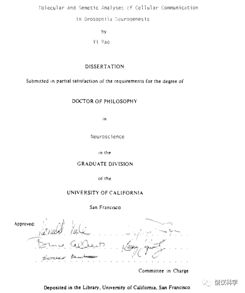
题目是
果蝇神经发生过程中细胞通讯的分子和遗传分析
Molecular and Genetic Analyses of Cellular Communication in Drosophila Neurogenesis
题目概括了论文研究使用的分子生物学和遗传学,动物,细胞机理,神经系统的发育。
突出了交叉:分子生物学,遗传学,神经生物学,发育生物学,细胞生物学。
在很长时间,这五个学科是我和我后来实验室主要的学科。
我讲课的内容也长期与这五个学科有关。
在北大之后,实验室逐渐加了:人类遗传学、化学分析、电生理、基因组学和成像。
论文委员会签名:Ronald Vale,Yuh-Nung Jan,Bruce Alberts,Larry Zipursky, Louis Reichardt(导师Jan)。
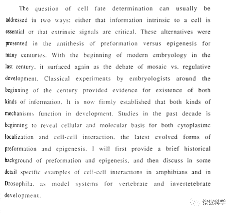
为了让引言言之有物,参考了从亚里士多德以来发育生物学重要文献,和发育遗传经典。从此一发不可收拾,后来经常是文献涵盖面比较广。
后来的讲课材料 《生物学概念与途径》可以说思路起源于当年的博士论文。
教材第五章的很多(和第六章小部分)参考了1991年的博士论文。翻译回英文,会有大段雷同。
1991年论文引用文献虽然是外国人为主,但也引用了三位华人的文章:李汝祺、童第周、许田。另外,在正文中提到庄孝惠的工作。
附1节选自《生物学概念与途径》第五章(比论文更详细)
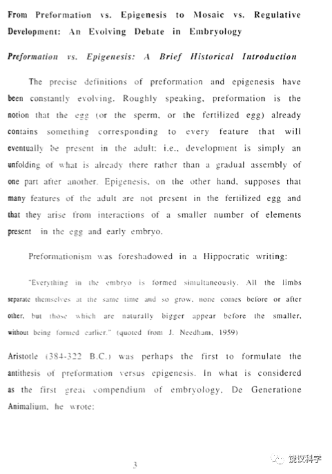
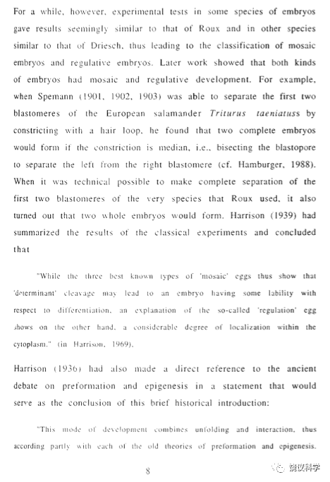
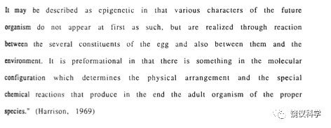
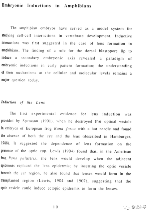
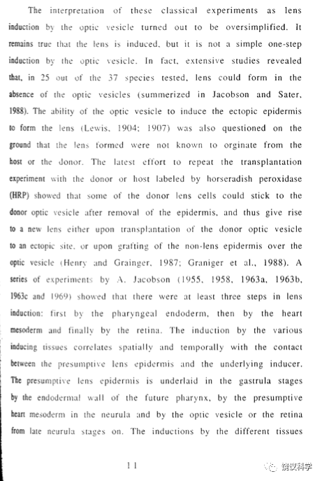
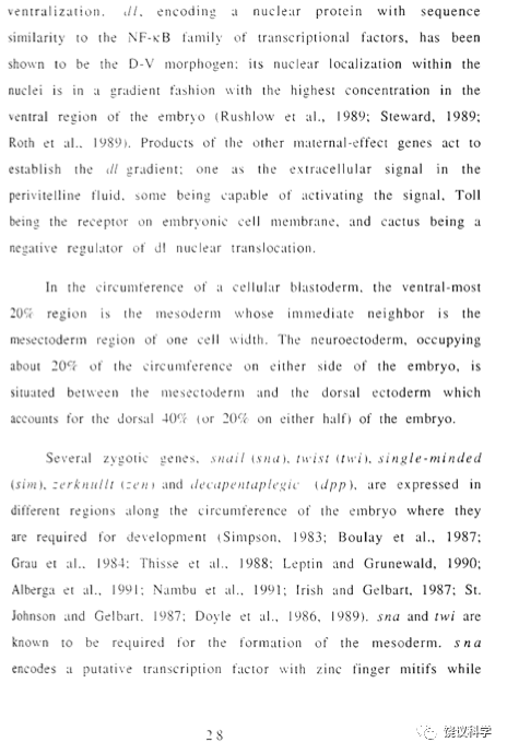
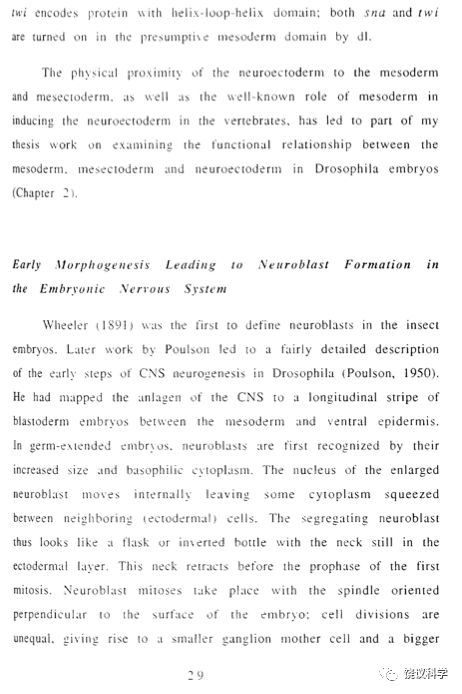
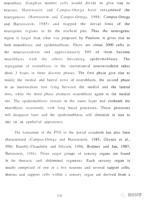
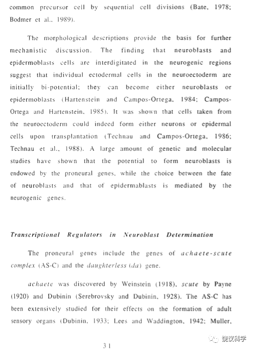
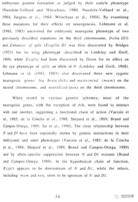
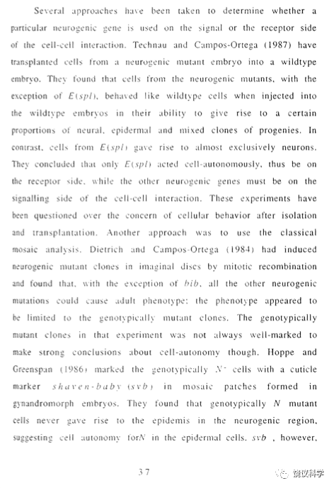
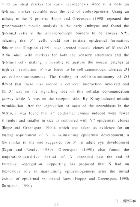
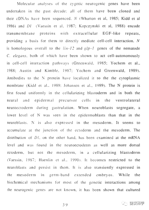
附2来自论文的拍照
附3引用文献来自1991年论文
附1节选自《生物学概念与途径》第五章
5 细胞间相互作用:胚胎诱导
发育生物学是研究一个细胞如何变成一个成体,并进一步变化直到死亡,包括细胞分裂、分化、生长、存活,组织和器官的形成和再生,个体的发育和衰老。发育生物学以前主要是研究胚胎发育,现在也研究超出胚胎基础过程的干细胞。而常为人类关心的组织、器官再生问题,缺乏基础研究的突破、有应用价值的结果更是微乎其微。发育生物学研究方法以前为简单的切割等操作,现代更多用分子生物学、也与物理和化学交叉。
5.1 预成论与后成论
动物的胚胎如何发育,曾有预成论(preformation)与后成论(epigenesis)的长期争论。预成论认为卵细胞(或精子、或受精卵)从开始就已形成了动物的各个部分,发育只是展开已有结构;而后成论认为受精卵并无成年的结构,而是在发育过程中逐渐形成。
希波克拉底(Hippocrates of Cos,公元前460-370)曾有类似预成论的观点:“胚胎中所有的都是同时形成。所有的肢体同时相互分开并生长,无一在其他之前或之后,而是那些自然较大的出现于较小的之前,并非更早形成”(引自Needham,1959)。
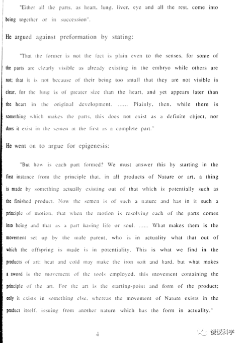
亚里士多德(Aristotle,公元前384-322)将预成与后成作为解释胚胎发育的两种对立概念。在《动物形成》一书中,他写道:“要么所有身体部件,如心、肺、肝、眼及其它,皆同时形成、或逐渐形成”。亚里士多德反对预成论:“前者不对是显而易见的,因为有些部件在胚胎已经清晰可见,而有些并非如此,且不是因为它们太小,比如肺比心大,但其发育出现晚于心……,那么就很明显,虽然有些东西制造部件,但非确定的物体,也不在精子中完整存在”。他支持后成论:“原则上,自然界一件东西来源于另外一件潜在可以成为终产物的东西……”。
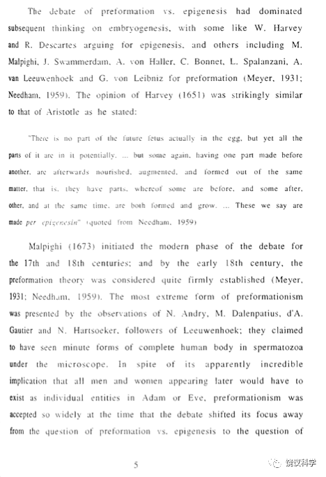
后成论的支持者包括英国的哈维(William Harvey,1578-1657)、法国的笛卡尔(Rene Descartes,1596-1650)等,预成论的支持者包括意大利的Marcello Malpighi(1628-1694)、荷兰的列文胡克(Antonie van Leeuwenhoek,1632-1723)、德国的莱布尼茨(Gottfried Wilhelm von Leibniz,1646-1716)等。哈维在1651年称:卵中并无未来胚胎的任何部件,但潜在含有所有部件…有些部件先形成、有些后形成,都继续生长…这样我们称为后成(Needham,1959)。Malpighi于1673年再启争论,到18世纪预成论被认为定论(Meyer,1931;Needham,1959)。荷兰显微镜专家Nicolaas Hartsoeker(1656-1725)设想如果在显微镜下可以看到精子显示人体所有部分,可能的画面如图2。此图在被误认为是他的观察、而非其设想的情况下广为传播(Hill, 1985)。如果精子就有整个人体,那么发育不过是长大,而无需形成新的结构,甚至早先的人(比如传说的亚当或夏娃)应该含有所有现代人类的个体。当时预成论广为接受,争论是预成于卵子、还是精子。
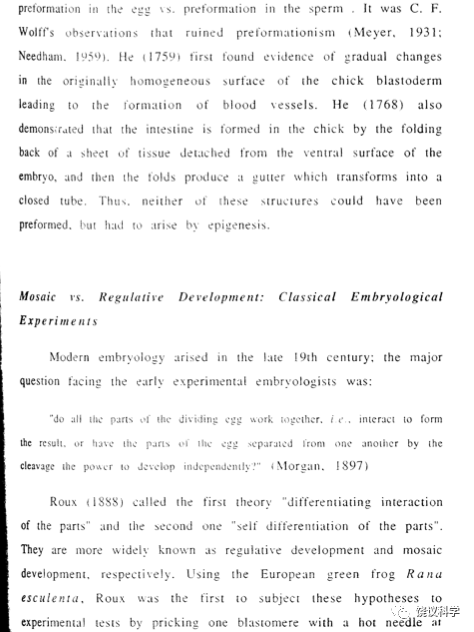
德国的Caspar Friedrich Wolff(1733-1794)观察鸡胚发育。他于1759年在“发生理论”的论文中重提后成论,并叙述鸡胚在原本匀质的表面逐渐形成血管,1768年在“小肠形成”一书中描述鸡胚的肠子从一片组织折叠形成,所以血管和小肠皆非预先形成,而是逐渐形成。Wolff的结果推翻了传统的预成论(Meyer,1931;Needham,1959)。
5.2 自分化与依赖性分化
十九世纪后期,德国科学家Wilhelm Roux(1850-1924)区分两种观点:“不同部分的自分化”、“不同部分的分化性相互作用”。
Roux称自己的研究为“发育成因学”(Entwicklungsmechanik)(Sander,1991a)。以前的科学家包括他的老师Ernst Haeckel(1834-1919),对胚胎的研究依赖于观察,在描述的基础上提出理论;而德国的Oskar Hertwig(1849-1922)、法国的Laurent Chabry(1855-1893)等开始了实验研究(Fischer, 1990; Sander,1991a);Roux提倡分析性实验,不仅要有实验结果、还要思考结果、检验理论(Sander,1991a;Hamburger, 1997)。他们开创的学科到二十世纪被称为“实验胚胎学”(Experimental Embryology)。
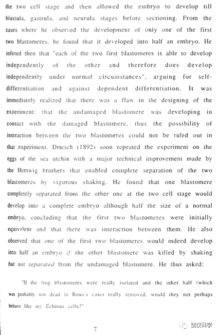
Roux选择了重要“自分化”还是“依赖性”分化作为研究的问题,实验模型用两栖类胚胎。1888年,他发表了对蛙胚研究的结果和结论。他集中研究受精卵第一次分裂后成为的两个细胞是自主发育、还是相互依赖。为此,他在受精卵分裂后,用加热的针“杀死”其中一个细胞,观察未受针刺的细胞如何发育,他观察到剩下的细胞本身继续分裂、发育后形成半个胚胎(图3),据此Roux认为最初两个细胞之一的失去并不影响另外一个细胞的发育,所以两个细胞发育是独立的(Roux,1888)。
德国的Hans Driesch (1867-1941)曾于1887年和1889年师从Haeckel。为了验证Roux的结论,Driesch在意大利Naples海洋生物实验站用海胆胚胎做实验。此前已知摇晃海胆的受精卵可以出现脱落的情况,Driesch在海胆受精卵分裂到两个细胞的时期,摇晃海胆几分钟,有些海胆胚胎的两个细胞会分开,Driesch将它们分别培养,然后观察分开的两个细胞如何发育。与Roux结论的预计不同,分开的两个细胞分别形成两个完整的、虽然体积只有正常一半大小的胚胎。如果在卵受精后分裂两次变成四个细胞时再摇晃,四个细胞都分别独立形成完整的胚胎(Driesch,1891)(图4)。这些结果显示了“调节”现象:胚胎的部分可以变成整体。对调节的解释说明:在胚胎两细胞期(或四细胞期),每个细胞都具有形成完整胚胎的潜力,但它们的相互作用导致每个细胞在其他细胞存在的情况下分别只参与形成半个胚胎(或四分之一个胚胎)。也就是说,细胞之间一定有相互作用。
虽然Roux未接受Driesch结果的重要性,认为低等的无脊椎动物海胆所得到的结论不如脊椎动物可靠,Roux和Driesch等创造了发育生物学的第一个高峰。Roux启迪了思路、开创了途径,Driesch有更严格的实验和分析。
德国的Hertwig(1893)最早试图用丝线分开两细胞期蛙胚的两个细胞(constriction,结扎实验),未成功(Beetschen, 1996)。美国的Morgan(1895)、意大利的Herlitzka(1895)、德国的Endres (1896)等曾有类似工作。
德国的Hans Spemann(1869-1941)在Würzburg大学随Theodor Boveri做研究生,1895年获博士学位。Spemann在Hertwig、Endres等基础上,在蝾螈受精卵第一次分裂后,用头发把两个细胞分开,结果两个细胞可以分别独立地形成完整的蝌蚪,证明脊椎动物胚胎的细胞也有相互作用(Spemann,1901,1902,1903)(图5左)。
Spemann发现结扎是否导致两个完整胚胎形成取决于胚胎第一次细胞分裂的平面(图5中和右)。如果分裂平面与胚胎未来的左右相应,分开的两个皆完整胚胎。如果分裂平面与背腹相应时,背侧部分可以形成一个几乎完整的胚胎(含神经系统等重要结构)、而腹侧部分只能形成皮肤等结构而不形成完整的胚胎,说明胚胎具有腹侧和背侧的不对称性。
Spemann认为腹侧缺乏某种“分化物质”:这种分化物质可能是器官形成或释放所需的未组织的物质、也可能是有组织的胚胎物质具有分化为各种器官的能力、或者引导其它细胞分化(Spemann,1901;Hamburger,1988)。因此,在这些可能包括胚胎背侧含有诱导其他细胞分化物质的想法的雏形。
5.3 晶状体诱导
器官中不同部件优美地配合,既是进化的奇妙、也是发育的奥秘。不同部件是否在发育中有相互关系?
例如:美妙的眼睛中,晶状体(lens)在解剖上位于视网膜前特定部位、功能上调节光线准确地投射到视网膜。那么在发育过程中,晶状体与视网膜的发育前体结构视杯(optic cup,亦称optic vesicle)是否相关?比如空间、时间的协调,抑或有依赖关系?
1901至1912年,Spemann曾研究晶状体形成,认为在发育早期,视杯诱导晶状体产生。支持诱导机理的实验包括:Spemann于二十世纪初报道在去除视杯后,晶状体不能形成(Spemann,1901b);美国霍普金斯大学的Warren Lewis(1870-1964)发现将原本不形成晶状体的外胚层移到视杯上方,可以形成晶状体(Lewis,1904);Lewis还发现将视杯移植到其他部位,导致新部位形成晶状体(Lewis,1906)(图6)。
其他研究者以及Spemann本人还看到有些胚胎晶状体产生不依赖视杯,当时以为有晶状体依赖视杯、和晶状体不依赖视杯的两类胚胎。1980年代后,视杯诱导晶状体的简单方式不被认可,而更复杂(Grainger et al., 1988)。
对于Spemann时代的胚胎学家来说,晶状体的研究为胚胎诱导的研究埋下了伏笔。
5.4 移植实验
两栖类胚胎发育最初从一个受精卵、分裂到几千个细胞皆为裂殖胚(blastula,所含的每个细胞称为blastomeres),其后肠胚(gastrula)。在动物极与植物极交界的附近,细胞开始内卷(invagination),为肠胚形成的开始。内卷首先从背侧开始、继而侧面、后在腹侧,最后形成一圈,为裂殖孔(blastopore),背侧内卷时偏动物极的上方部位称为“背唇”(the dorsal blastopore lip,简称the dorsal lip)(图7)。
十九世纪Roux与Driesch等实验对象为裂殖胚。Spemann将结扎实验延伸到不同的胚胎发育期,发现一直到肠胚期如果分开背腹部分都有差别。分离的腹侧部只能形成皮肤、和腹侧及侧面中胚层组织如血液和间充质等,而分离的背侧部产生神经系统、和背侧的中胚层结构如脊索和体节。在分离时,背侧部分含背唇。
Roux和Morgan认为胚胎早期赤道部分对发育很重要,Roux还认为背唇可以产生背侧的外胚层结构神经管(Roux, 1892; Morgan, 1894; Lewis, 1907)。
1907年,霍普金斯大学的Lewis将肠胚期的背唇从一个供体胚胎移植到发育更晚期的宿主胚胎,结果发现宿主胚胎因为移植而多出现背侧中胚层组织脊索和肌肉、及外胚层组织神经管(图8)。Lewis认为:来自供体胚胎的背唇被移植到宿主的后自主形成了中胚层和外胚层组织,因为背唇的发育命运已经决定了(determined)(Lewis,1907),也就是认为背唇自主发育为脊索、肌肉和神经管,没有提出其中是否有因为背唇诱导宿主而产生的结构。
1916年,Spemann利用常见蝾螈(Triturus taeniatus)每个胚胎着色的自然差别,在两个同种蝾螈胚胎之间进行移植,通过着色深浅跟踪移植的供体在宿主胚胎中的发育命运。他发现肠胚期神经前体区域如果移植到另一胚胎的表皮区域,会形成表皮,而肠胚的表皮区域移植到神经区域会形成神经,都不按供体原有命运,而按被移植到的宿主区域改变了发育命运,说明在移植前它们的命运没有固定,而可以为新邻居所影响再确定(Spemann,1916,1918,1919)。
Spemann用蝾螈重复了Lewis蛙的背唇移植实验,获得两根背轴。最初记录在他1916年4月20日的笔记中(Fäbler,1996),1918年发表两根背轴分别含神经管、脊髓(图9)(Spemann,1918)。
但Spemann与Lewis一样,以为供体的背唇直接成为脊索和神经组织,而未意识到出现了诱导现象。Spemann在1918年的文章中还标明第二根神经管来源于移植的背唇(Spemann, 1918;Sander and Fässler,2001)(图10)。
令人惊讶的是,虽然Lewis和Spemann都曾积极研究过晶状体诱导,试图用不同方法证明或否定晶状体是否依赖于视杯,他们两人皆未考虑第二轴中是否有组织是因为供体诱导宿主而产生。这在Spemann尤为突出,因为他在Lewis文章九年后发重复性质的文章,而且获得的第二轴很漂亮。他还是与Lewis一样以为背唇成为脊索和神经管。在他1918年文章中,他标注第二根神经管来源于背唇(图10)(Spemann, 1918; Sander and Fässler, 2001)。Spemann的错误来源于对背唇命运的错误认识。1892年,Roux 的结论是“依据于三种不同的实验方法,…背唇的外表面是神经管的位置”(Roux, 1892; Sander, 1991c)。Spemann因此认为肠胚期的背唇的深层和浅层分别成为脊索和神经管(图11)(Hamburger, 1988; Fäbler,1996),他称背唇为“分化中心”(Spemann, 1918)。
如果Spemann仔细分析自己在1918年发表的结果(图12),可以看到:神经管来源的区域在背唇以上的区域。
1919年海德堡大学的Hans Petersen(1885-1946)在读到Spemann(1918)后,去信Spemann指出背唇的表层也应该是中胚层(脊索、体节)、而不形成神经管(Spemann, 1921; Hamburger,1988;Sander and Fässler,2001)。因此Spemann决定要研究背唇移植后导致的第二根背轴中脊索和神经管到底是来自移植供体、还是宿主。
Spemann于1916和1918年报道的移植是在同种胚胎之间,依赖供体和宿主同种的两个个体之间自然的色素差异,在分析时不太容易区分。为了确切区分背唇的命运,需要在颜色不同的不同种胚胎之间做背唇移植。1896年,德国犹太科学家Gustav Jacob Born(1852-1900)首先在异种胚胎间移植,他从一种蛙胚切下一块移植到另一胚胎上形成嵌合胚胎(chimera)(Born,1896)。1898年,美国霍普金斯大学的Ross Harrison (1870-1959)用同样方法在不同种、不同色素的蛙之间进行过胚胎组织的移植(Harrison,1898),可以在移植后发育成的蝌蚪中区分来源于供体和宿主的组织。Spemann于1916年做同种移植(homeoplastic transplantation)、1917年开始做异种移植(heteroplastic transplantation)。Spemann用不同颜色的蝾螈之间进行移植,他常用色浅的Triturus cristatus和色深的Triturus taeniatus(或Triturus alpestris),以便区分供体和宿主来源的胚胎组织。
Hilde Mangold(1898-1924)上大学主修自然科学,她在Frankfurt大学听Spemann的学术报告后要求转学到Spemann所在的Freiburg大学,1920年进Spemann实验室做研究生(Fässler and Sander,1996)。Spemann给她的第一个课题是研究水螅(hydra),但实验遇到问题、Spemann也不能解决,再给她的课题是Spemann自己做过、所以技术上应该没有问题:背唇移植。将来源于色浅的T cristatus的背唇移植到色深的T taeniatus(或T alpestris)。
在Mangold尚未开始移植实验的1921年1月,Spemann单独提交的文章写道:“也许整个移植的背唇属于…内中胚层…如果这样的话,从taeniatus胚胎来的背唇移植到cristatus宿主后,(中胚层的)脊索和体节应该属于前者,而(外胚层)的神经板属于后者”(Spemann,1921;Sander and Fässler,2001)。
很可能是在初稿提交后Mangold的移植实验开始有了结果,Spemann将Mangold的一个结果于1921年5月作为后记补录在Spemann(1921)论文中(Spemann,1921;Sander and Fässler,2001)。Mangold将色浅的T cristatus肠胚期背唇移植到色深的T taeniatus肠胚侧面,观察到第二个背轴产生。1921年,Spemann用了组织者(organizer)一词描述背唇:在它正常存在或者移植到的部位,对未分化材料创造一定方向和程度的“组织场”(Spemann,1921;Hamburger,1988)。
5.5 胚胎诱导与组织者
1921年和1922年,Mangold记录了259次移植实验,其中28个宿主胚胎出现第二背轴,当时培养胚胎的溶液欠佳、也无抗生素,所以胚胎在手术后的成活率不高。Hilde于1923年曾与丈夫Otto Mangold合作做移植实验(217个宿主,其中17个有第二背轴),Spemann没选Mangold夫妇合作获得的结果(Fässler and Sander,1996)。
1924年,Spemann和Mangold发表合作的文章,Spemann选择详细分析了Mangold获得的六个胚胎移植的结果(Spemann and Mangold,1924)。
文章首先指出蝾螈肠胚化开始时,不同部位的决定性不同,通过移植可以检验,发现有些组织移植到其他部位后,其发育命运可以改变,这些工作Spemann于1918和1921已报道,而为Otto Mangold于1922年、1923年所验证和推广。然后,他们指出,Spemann(1918)就发现背唇很不一样,移植到形成上皮的地方,背唇继续按自己确定的命运发育,而移植后的区域出现第二个胚胎原基,含神经管、脊索和体节。Lewis(1907)有类似的发现,只是用肠胚后期的背唇移植到更老一些的胚胎其他部位,观察到它发育为神经组织和体节。
Spemann和Mangold提出发育命运确定在先的区域可以决定其他尚未确定命运区域的发育。他们用的例子是胚胎旋转的实验结果。蛙卵细胞有极性,着色深、较轻的为动物极,着色浅、较重的为植物极(图15),这一极性维持到胚胎发育过程中,精子从动物极进入卵细胞,动物半球主要贡献于外胚层和中胚层,植物半球主要贡献于内胚层。1918年Spemann的研究中曾将肠胚的动物极半球、植物半球分开,然后180度或90度旋转后再重合,结果认为植物极影响动物极的发育。由此,他们用了“组织中心”(organization center)的概念:胚胎中先决定的区域,再送出一定方向和一定量的决定性影响。
他们认为虽然Spemann(1918)发现了胚胎不同区域的决定性不同,但当时只在同种胚胎之间进行移植,不易区分移植供体与宿主,而异种移植更好,比如将T cristatus胚胎中命运确定的区域移植到T taeniatus(或T alpestris)胚胎中未定区域。
文章描述的六个移植为1921年的Um8b,和1922年Um25b、Um214、Um131b、Um83b、Um132b。文章只在讨论Um25时有文字介绍、而未显示Um214的图片,而多个图片显示了其他每个移植胚胎在多个部位的切片。
Um8b第二根神经管中很长部分来源于宿主、但有相当部分来源于供体,脊索来自供体;Um25b第二根神经管完全来自于宿主,而脊索完全来源于供体;Um214b的第二根神经管也完全来自于宿主,而脊索完全来源于供体;Um131b的第二根神经管多来自宿主、少数部位细胞来自供体,脊索和体节来自供体;Um83b的第二根神经管来自宿主,体节多数细胞来自宿主,脊索和部分体节来自供体;Um132b第二根神经管多数来自宿主(后2/3部分少数细胞来自供体,皆在神经管的底板),脊索与部分体节来自供体(图13)。可能是因为Um25b的两张组织切片的图不特别好看,所以一般教科书不太用这一最能突出神经管可能由脊索诱导产生的例子,而常用Um132b为例,Um132的第二根神经管比较长,其胚胎在体外培养的时期也较长。
在讨论中,Spemann和Mangold先谈了移植的方向、背唇的具体位置、发育期、第二原基内涵等。在机理讨论部分,他们明确提出肯定出现了诱导,但不清楚发生的确切的时间、是否直接诱导,提出两种可能:1)移植供体本身产生少量神经组织并存在于外胚层表面(如Um8b所见),而这些神经组织诱导宿主的细胞成为神经组织;2)中胚层诱导神经组织产生。他们当时的结果不能确定哪种解释为正确,提出了一些可能的实验。他们还定义和讨论了组织者的概念。
什么是组织者、组织者如何起作用可能在今天还有争论,而诱导为后来很多实验所证明,并不限于胚胎、也不限于发育,是细胞之间相互作用的一种范式。
5.6 余音缭绕
Walther Vogt(1888-1941)从1920年代初开始用染料跟踪胚胎细胞的命运(Vogt, 1923),并于1925和1929年发表蛙胚的命运图(fate map)(Vogt, 1929)(图14)。他的图谱清楚地显示背唇本身只成为中胚层,Spemann以前以为背唇深层和浅层不同命运、浅层成为神经胚层的想法肯定不对。
Spemann的研究生Johannes Holtfreter(1901-1992)、Otto Mangold等推进了神经诱导的研究,包括诱导的区域性、建立体外诱导模型等。Holtfreter实验室的中国留学生庄孝惠研究了宿主胚胎不同区域对外源性诱导物的反应(Hamburger et al., 1988)。
1919至1924年Spemann没做实验,1925年重回实验第一线,1929年开始用不同方法“破坏”背唇的结构(切、压、加热、冷冻、干燥),观察经过这些处理后,背唇是否还有诱导的活性(Hamburger, 1988),结果多为阴性。Holtfreter离开Spemann实验室后独立做过类似的研究,他改进了培养液,大大提高了胚胎的存活率,他发现多种方法处理后的背唇仍然有神经诱导的活性。1932年,Spemann与三个学生合作发表了一篇论文、四人分别写一段(Bautzmann et al., 1932),其中Holtfreter一人做过130个胚胎,其他人做过少量的胚胎:Hermann Bautzmann观察到背唇加温到60度处理5到10分钟后,还可能有神经诱导活性;Holtfreter发现背唇在60度干燥、100度加温、冷冻后,仍有神经诱导活性;Spemann发现背唇用96%的酒精处理3分钟后,仍有诱导活性,不过他只有一例胚胎;Mangold希望看诱导物质是否可弥散,将背唇附着于琼脂,干燥后将琼脂转到胚胎,检验其诱导性。Bautzmann等(1932)证明无需活着的背唇组织或细胞就可能有诱导活性,被认为开启了胚胎诱导分子的研究。
Spemann的研究生Else Wehmeier与化学家Fischer合作,试图用丙酮、乙醇、乙醚等提取背唇的神经诱导物质(Fischer and Wehmeier,1933)。剑桥大学的李约瑟(Joseph Needham,1900-1995)、Dorothy Moyle Needham(1896-1987)和Conrad Waddington(1905-1975)研究蝾螈提取物的诱导活性(Waddington et al., 1933; Needham et al., 1934)。但是,生化胚胎学(biochemical embryology)热潮中,并未得到诱导的分子。
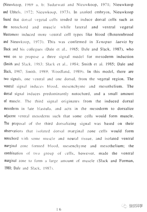
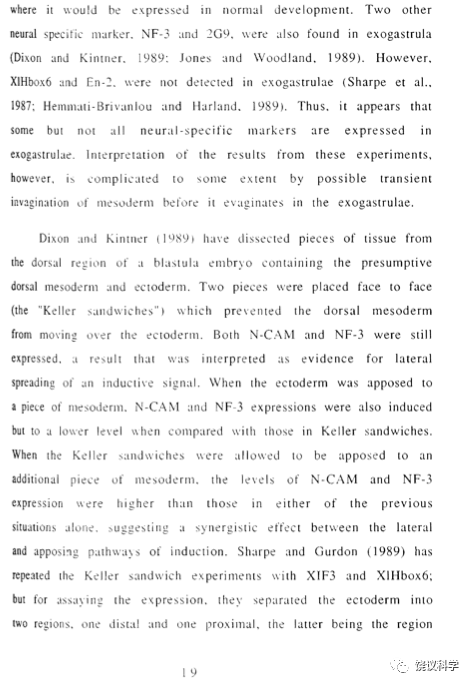
1940年代至1970年代的三十年为胚胎发育研究的寒冷期。其中最重要的是荷兰Utrecht大学的Pieter Nieuwkoop(1927-2007)于1969年发现中胚层为内胚层诱导所产生。Nieuwkoop将裂殖胚从动物极到植物极分成几个部分,发现虽然动物极单独不能形成中胚层,但它如果与植物极共同培养,可以形成中胚层(Nieukwoop,1969a, 1969b, 1970)。进一步通过同位素标记区分供体和宿主证明在植物极与动物极共培养时,中胚层来自动物极,所以是植物极诱导动物极产生中胚层(Nieukwoop and Ubbels,1972)。在正常发育过程中,中胚层之所以在动物极和植物极之间产生,是因为植物极发出信号诱导动物极和植物极之间的结构(Morgan时代称赤道区、后称边缘区marginal zone)形成中胚层。
由于背唇神经诱导曾被质问(Jacobson, 1982, 1983),英国两个实验室于1983年将辣根过氧化酶(horseradish peroxidase,HRP)注射到供体胚胎,在肠胚早期获得背唇,移植到同期宿主的腹侧,观察到第二根背轴,其中HRP只存在于脊索中,而神经管完全来源于宿主(Smith and Slack,1983;Gimlich and Cooke,1983),从而回答了质疑、再次证实了1924年的结论。
肠胚期胚胎的腹背轴已经形成,因此肠胚背唇移植导致背轴形成的实验需要在胚胎更早期进行研究。美国伯克利加州大学的John Gerhart实验室在64细胞期做移植实验,发现植物半球背侧的单个细胞如果移植到宿主胚胎的腹侧,可以诱导第二背轴产生(Gimlich and Gerhart,1984)。实际上,腹背轴的形成可以推早到受精:在受精前,卵细胞无腹背差别,而精子进入导致卵细胞内细胞骨架变化、引起一系列变化,致使精子进入一侧为将来胚胎的腹侧(Gerhartet al., 1989)。
所以,背唇移植实验延伸出中胚层诱导、腹背轴形成等多个重要问题的研究。
5.7 分子时代
1930年代没找到胚胎诱导相关的分子;1970年代重组DNA为代表的新技术导致分子生物学在生物医学多个领域的应用;1980至1990年代不同的方法帮助找到了中胚层诱导、背轴诱导和神经诱导的分子,并对神经诱导的机理有新的理解。
因为胚胎植物极有诱导中胚层的活性,哈佛大学的Douglas Melton通过mRNA差异性分布,找到非洲爪蟾卵细胞动物极和植物极分别特有(或富集)的mRNA(Rebagliatiet al., 1985),其中植物极特有的Vg-1编码蛋白质为转化生长因子(TGF-b)家族的成员(Weeks and Melton,1987)(图16)。美国旧金山加州大学(UCSF)的Kimelman和Kirschner用裂殖胚的动物极部分(所谓“动物帽”,animal cap)体外培养作为检测方法,筛查当时已知生长因子的诱导活性。动物帽单独培养只形成皮肤,而培养时如果存在成纤维细胞生长因子(fibroblast growth factor,FGF),可以出现中胚层(特别是腹侧和侧面的中胚层组织)(Kimelman and Kirschner,1987)。英国的Jim Smith发现爪蟾体外培养细胞系的分泌物有中胚层诱导活性(Smith, 1987),以后被证明为activin、也是TGF-b家族成员。Activin的作用有剂量依赖性,低剂量诱导腹侧、侧面中胚层,高剂量诱导背侧中胚层(如背唇、或其衍化的脊索)。TGF-b家族的骨形成蛋白(BMP,bone morphogenetic protein)可以诱导腹侧中胚层产生。
一个偶然的发现开启了对背轴形成分子机理的理解。研究癌基因的英裔美国分子生物学家Andrew McMahon与研究蛙胚的Randall Moon合作,将认为在老鼠过高或者异位表达导致乳腺癌的int-1基因所转录的mRNA注射到爪蟾受精卵,结果胚胎形成两个背轴(McMahan and Moon,1989),这一意外的发现当时被认为是“分裂”了组织者,其后认为是激活了背轴形成的通路。从研究病毒导致老鼠癌症发现的int-1与从研究果蝇翅膀形成发现的wingless基因组成Wnt家族,产生分泌型蛋白,Wnt不同成员通过作用于其受体及信号转导分子参与多个生物过程。它们在蛙胚参与背轴形成,但尚待确定胚胎内从哪一步开始用Wnt信号通路。1992年,伯克利加州大学的英裔美国科学家Richard Harland实验室发现表达于背唇的Noggin基因,编码分泌型蛋白质也能诱导背轴(Smith and Harland,1992)。
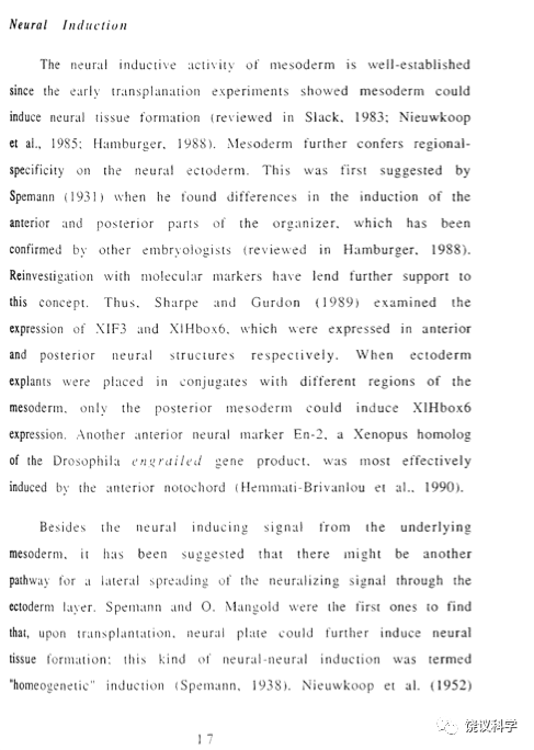
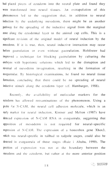
1993年,Harland实验室发现Noggin有神经诱导的活性(Lamb et al., 1993)。1994年,洛杉矶加州大学的阿根廷裔美国科学家De Robertis实验室发现背唇表达Chordin、也编码有神经诱导活性的分泌型蛋白质(Sasai et al., 1994)(图17)。
Melton实验室的伊朗库尔德裔Ali Hemmati-Brivanlou在研究activin参与中胚层诱导的过程中,用activin受体制造的抑制分子阻止中胚层形成时,意外发现没有中胚层仍可出现神经系统,从而提出神经胚层的诱导在分子机理上是去抑制:胚胎内存在抑制神经胚层形成的分子,而如果抑制这些抑制性分子就可以“诱导”神经发生(Hemmati-Brivanlou and Melton, 1994)。抑制分子可能是BMP,而noggin和chordin等蛋白质可以直接结合BMP、并抑制BMP的活性(Piccoloet al., 1996;Zimmermanet al., 1996)。
5.8 结语
预成论与后成论的古老争论,在实验胚胎学兴起后,刺激了人们感兴趣研究早期细胞发育是完全独立自主、还是相互作用。
Spemann和Mangold(1924)的研究,实验设计类似于Lewis(1907)。Spemann曾于1916年、1918年重复Lewis的工作并沿用其理解,而Petersen于1919年向Spemann提出以前对背唇自分化命运的理解可能有问题,加上概念上曾有Spemann和Lewis等在二十世纪初研究晶状体可能被诱导产生的铺垫,导致Spemann决定采用Born(1896)和Harrison(1898)的方法,通过异种胚胎间移植确切地分清移植提供的背唇自身变成神经管、还是诱导宿主胚胎组织产生神经管,最终Spemann指导Mangold的实验获得结果提示神经管为背唇诱导所产生。
胚胎诱导的概念,不仅成为核心问题之一主导几十年的发育生物学研究,也影响了人们思考细胞相互作用所参与的其他生物学过程,但1930年代研究胚胎诱导的分子机理遭遇技术困难,直到1980、1990年代才获突破。
在实验胚胎学研究过程中多次发现不同方法可以产生连体胚胎,包括结扎实验、背唇移植实验、早期裂殖胚细胞移植实验、Wnt家族分子注射、以及本文未讨论的BMP受体抑制剂注射。这些研究帮助我们理解基本的发育生物学机理,但是,迄今并不清楚连体人的发生原因。
附2来自论文的拍照

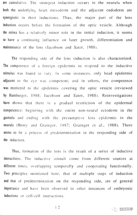
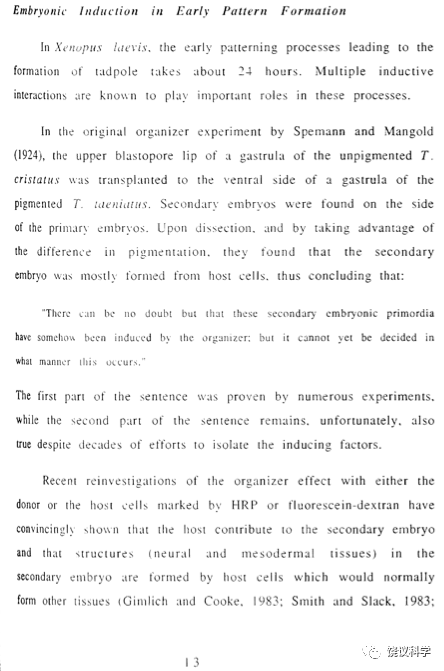
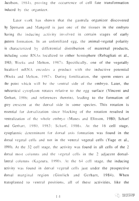
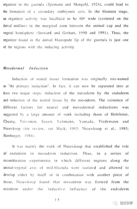
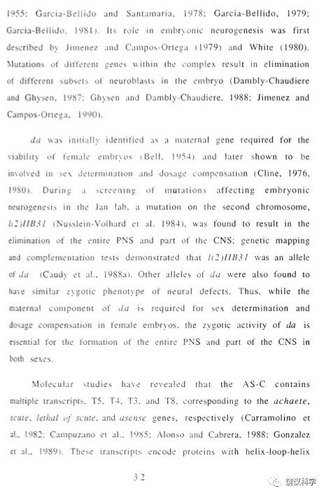
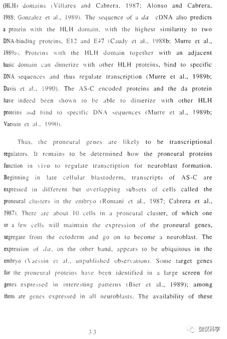
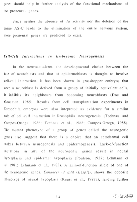
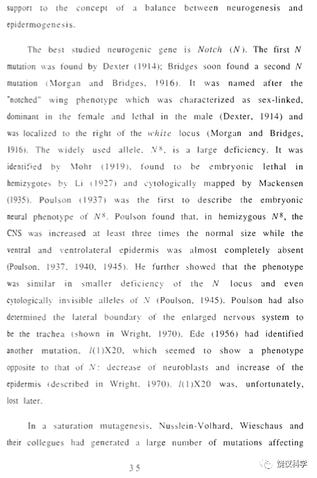
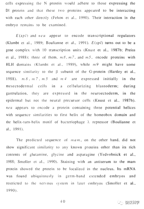
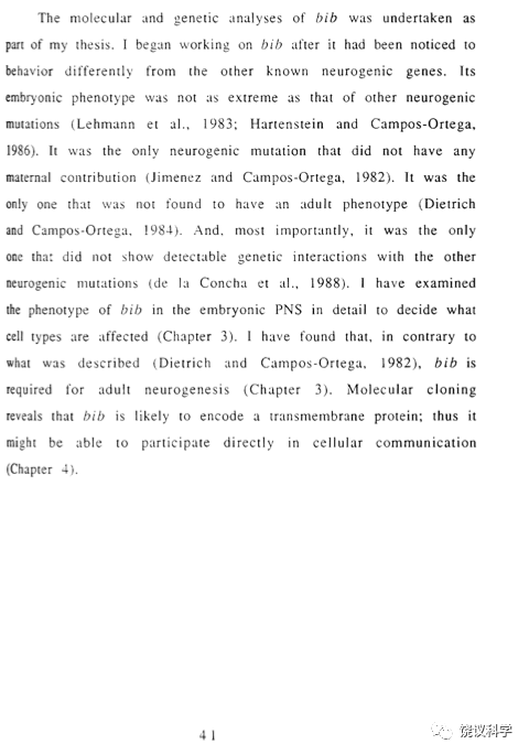
附3引用文献来自1991年论文
Alberga, A., Boulay, L-L., Kempe, E., Dennefe1d, C. and Haenlin, M. (1991). The snail gene rewuired for mesoderm formation in Drosophila is expressed dynamically in derivatives of all three germ layers. Development 111:983-92
Alonso, M.C. and Cabrera, C.V.(1988).The achaete-scute gene complex of Drosophila melanogaster comprises four homologous genes.EMBO J.7:2585-91
Amaya,E., Musci, T.J. and Kirschner, M.(1991). Expression of a dominant negative mutant of the FGF receptor disrupts mesoderm formation in Xenopus embryos. Cell, in press
Anderson, K.V. (1987). Dorsal-ventral embryonic pattern genes of Drosophila. Trends Genet.3:91-97
Anderson, K.V. and Nusslein-Volhard, C. (1984). Information for the dorsal-ventral pattern of the Drosophila embryo is stored as maternal mRNA. Nature 311:223-7
Anderson, K.V. and Nusslein-Volhard,C. (1985). Establishment of dorsal-ventral polarity in the Drosophila embryo: the induction of polarity by the Toll gene product. Cell 42:7 91-8 9
Aristotle (384-322, B. C.). De Generatione Animalium, translated by A. Platt, (Oxford: Clarendon Press, 1910)
Artavanis-Tsakonas, S. (1988). The molecular biology of the Notch locus and the fine tuning of differentiation in Drosophila. Trends Genet.4:95-100
Austin, J., and Kimble, J. (1987). glp-l is required in the germ line for regulation of the decision between mitosis and meiosis in C. elegans. Cell 51:589-99
Bate, C.M. (1978). Development of sensory systems in arthropods. In Handbook of Sensory Physiology, ed M.Jacobson, 9:1 -53. (Berlin:S pringer-Ve lag)
Bell, A.E. (1954). A gene in Drosophila melanogaster that produces all male progeny. Genetics 39:958-9
Bier, E., Ackerman, L., Barbel, S., Jan, L., and Jan, Y.N. (1988). Identification and characterization of a neuron-specific nuclear antigen in Drosophila. Science 240:913 916
Bier, E., Jan, L.Y. and Jan, Y.N.(1990). rhomboid,a gene required for dorso-ventral axis establishment and peripheral nervous system development in Drosophila melanogaster. Genes Dev.4:190-203
Blochlinger, K.B, Bodmer, R., Jack, J., Jan, L.Y. and Jan, Y.N.(1988). Primary structure and expression of a product from cut, a locus involved in specifying sensory organ identity in Drosophila. Nature 333:629-635,
Blochlinger. K.B., Bodmer,R., Jan, L.Y. and Jan, Y.N. (1990). Patterns of expression of cut, a protein required for external sensory organ development in wild-type and cut mutant Drosophila embryos. Genes Dev. 4:1322-1331
Bodmer, R., Barbel, S.. Shepard, S., Jack, J., Jan, L.Y. and Jan, Y.N. (1987).Transformation of sensory organs by mutations of the cut locus of D. melanogaster. Cell 51:293-307
Bodmer, R. and Jan,Y.N. (1987). Morphological differentiation of the embryonic peripheral neurons in Drosophila. Raux's Arch. Dev. Biol. 196:69-77
Bodmer, R., Carretto, R. and Jan, Y.N. (1989). Neurogenesis of the peripheral nervous system in Drosophila embryos: DNA replication patterns and cell lineages. Neuron 3:31 -7
Bodmer, R., Jan.L.Y.and Jan, Y.N.(1990).A new homeobox- containing gene,msh-2,is transiently expressed early during mesoderm formation of Drosophila Development 110:661-9
Bok, D., Dockstader. D. and Horwitz, J. (1983). Immunocytochemical localization of the lens main intrinsic polypeptide (MIP) in communicating junctions. J.Cell Biol.97:1491-1499
Boterenbrood, E.C. and Nieuwkoop, P.O. (1973). The formation of the mesoderm in urodelean amphibians. V. Its regional induction by the endoderm. Roux' Arch. Dev. Biol.173:319-32
Boulay, J.L., Dennefeld, C., and Alberga, A. (1987).The Drosophila developmental gene snail encodes a protein with nucleic acid binding fingers. Nature 330:395-398
Brand, M. and Campos-Ortega, J.A. (1988). Two groups of interrelated genes regulate early neurogenesis in Drosophila melanogaster. Roux's Arch. Dev. Biol.197:457-470
Brand, M. and Campos-Ortega, J.A. (1989). Second site modifiers of the split mutation of Notch define genes involved in neurogenesis in Drosophila melanogaster. Roux's Arch. Dev. Biol. 198:275-85
Broekhuyse, R.M., Kuhlman, E.D. and Winkens, H.J. (1979). Lens membranes. VII.MIP is an immunologically specific component of lens fiber membranes and is identical with 26K band protein. Exp. Eye Res.29:303-313
Brown, N.H. and Kafatos, F.C. (1988). Functional cDNA libraries from Drosophila Embryos. J. Mol. Biol. 203:425-437
Cabrera, C.V. (1990). Lateral inhibition and cell fate during neurogenesis in Drosophila: the interaction between scute, Notch and Delta. Development 109:733-42
Cabrera, C.V., Martinez-Arias, A., and Bate, M. (1987). The expression of three members of the achaete-scute gene complex correlates with neuroblast segregation in Drosophila. Cell 50:425-433
Cagan, R.L. and Ready, D.F. (1989). Notch is required for successive cell decisions in the developing Drosophila retina. Genes Dev .3:1099-1112
Campos-Ortega, J.A. (1983). Topological specificity of phenotype expression of neurogenic mutations in Drosophila . Roux's Arch. Dev. Biol. 192:317-326
Campos-Ortega, J.A. and Hartenstein, V. (1985). The Embryonic Development of Drosophila melanogaster.(Berlin, Heidelberg: Springer Verlag).
Campos-Ortega, J.A. and Jan, Y.N. (1991). Genetic and molecular bases of neurogeneis in Drosophila melanogaster. Annu. Rev. Neurosci. 14:399 420
Campuzano, S., Carramolina, L., Cabrera, C.V., Ruiz-Gomez, M., Villares, R., Boronat, A. and Modolell, J. (1985). Molecular genetics of the achaete-scute gene complex of Drosophila melanogaster. Cell 40:327-338
Carramolino, L., Ruiz-Gomez, M, Guerrero, M.C., Campuzano, S. and Modolell, J. (1982). DNA map of mutations at the scute locus of Drosophila melanogaster.EMBO J.1:1185-91.
Caudy, M., Grell, E.H., Dambly-Chaudiere, C., Ghysen, A, Jan, L. Y. and Jan, Y.N. (1988a).The maternal sex determination gene daughterless has zygotic activity necessary for the formation of peripheral neurons in Drosophila. Genes Dev.2:843-52
Caudy, M.. Vaessin, H., Brand, M.,Tuma,R., Jan, L.Y. and Jan, Y.N. (l988b). daughterless,a Drosophila gene essential for both neurogenesis and sex determination,has sequence similarities to myc and the aclzaete-scute complex. Cell 55:1061-1067
Cavener, D.R. (1987). Comparison of the consensus sequence flanking translational start sites in Drosophila and vertebrates. Nucl.Acids Res.15:1353-1361
Christian. J.L., Gavie, B.I., McMahon. A.P. and Moon, R.T. (1991a) Isolation of cDNAs partially encoding four Xenopus Wnt-l/int-l- related proteins and characterization of their transient expression during embryonic development. Dev. Biol. 143:230-234
Christian, J.L., McMahon, I.A., McMahon, A.P. and Moon, R.T. (1991b) Xwnt-8, a Xenopus Wnt-l/int-l-related gene responsive to mesoderm-inducing growth factors, may play a role in ventral mesodermal patterning during embryogenesis. Development 111:1045-1455
Cline,T.W. (1976). A sex-specific, temperature sensitive maternal effect of the daughterless mutation of Drosophila melanogaster. Genetics 84:723-42
Cline, T.W. (1980). Maternal and zygotic sex-specific gene interactions in Drosophila. Genetics 96:903 -26
Cooley, L.K.. Kelley,R. and Spradling, A. (1988).Insertional mutagenesis of the Drosophila genome with single P elements. Science 239:1121-1128
Dale, L. and Slack, J.M.W. (1987). Regional specification within the mesoderm of earlv embryos of Xenopus laevis. Development 100:279-95
Dale, L., Smith,J.C. and Slack,J.M.V.(1985).Medoserm induction in Xenopus laevis:a quantitative study using a cell lineage label and tissue-specific antibodies.1.Embryol.expo A1orph.89:289-312
Dambly-Chaudiere, C., and Ghysen, A. (1987). Independent subpatterns of sense organs require independent genes of the achaete-scute complex in Drosophila larvae. Genes Dev.1:297-306
Dawid, L B. and Sargent, T.D. (1988). X. laevis in developmental and molecular biology. Science 240:1443-1448
Davis. R.L., Cheng, P.-F., Lassar, A.B. and Weintraub, H. (1990). The MyoD DNA binding domain contains a recognition code for muscle- specific gene activation. Cell 60:733
de la Concha, A., Dietrich, U., Weigel, D., and Campos-Ortega, J.A. (1988). Functional interactions of neurogenic genes of Drosophila melanogaster. Genetics 118:499-508
Dexter, J.S. (1914). The analysis of a case of continuous variation in Drosophila by a study of its linkage relations.Amer.Nat. 48:712-58
Dixon, J.E. and Kintner, C.R. (1989). Cellular contacts required for neural induction in Xenopus embryos: evidence for two signals. Development 106:749-757
Doe, C.Q., and Goodman, C.S. (1985). Early events in insect neurogenesis. II. The role of cell interactions and cell lineages in the determination of neuronal precursor cells. Dev. Biol. Ill:206-219
Doyle, H.J., Harding, K., Hoey, T. and Levine,M (1986). Transcripts encoded by a homeobox gene are restricted to dorsal tissues of Drosophila embryos. Nature 323:76-9
Doyle, H.J., Kraut, R. and Levine,M (1989). Spatial regulation of zenknullt: a dorsal-ventral patterning gene in Drosophila. Genes Dev. 3:1515-33
Driesch, H. (1892). The potency of the first two cleavage cells in echinoderm development. Experimental production of partial and double formations. Zoologie 53:160-178, translated by Mezger, L., Hamburger, M. and Y., and Hall, T.S. in Willier, B.H. and Oppenheimer.J.M. ed. Foundations of Experimental Embryology (Prentice-Hall, Inc, Englewood Cliffs, N.J.) (1964)
Dubinin, N.P. (1933). Step allelomorphism in Drosophila melanogasler. J. Genetics 27:443-64
Ellis, H.M..Spann. D.R. and Posakony, J.W. (1990). extramacrochaetae. a negative regulator of sensory organ development in Drosophila, defines a new class of helix-loop-helix proteins. Cell 61:27-38
Ehring,G.R., Zampighi, G.A. and Hall, I.E. (1988). Properties of MIP 26 channels reconstituted into planar lipid bilayers. In:Gap Junctions, (eds E.L.Hertzberg&R.G.Johnson), pp.335-346, (New York:Alan R. Liss)
Fehon, R.G., Kooh,P .J., Rebay, I., Regan, C.L., Xu, T., Muskavitch, M.A.T. and Artavanis-Tsakonas, S. (1990). Molecular interactions between the protein products of the neurogenic loci Notch and Delta, Two EGF-homologous genes in Drosophila. Cell 61:523-34
Fortin, M.G., Morrison, N.A. and Verma, D.P.S.(1987). Nodulin-26, a peribacteroid membrane nodulin is expressed independently of the development of the peribacteroid compartment. Nucl. Acids Res.15:813-824
Garcia-Bellido, A. (1979). Genetic analysis of the achaete-scute system of Drosophila melanogaster. Genetics 91:491-520
Garcia-Bellido, A. (1981). From the gene to the pattern: Chaeta differentiation.in Lloyd C.W.and Rees, D.A.,eds, Cellular Controls in Differentiation. pp281 304, Acdemic Press
Garcia-Bellido, A. and Santamaria,P. (1978). Developmental analysis of the achaete-scute system of Drosophila melanogaster. Genetics 88:469-86
Garrell, J, and Modolell, I. (1990). The Drosophila extramacrochaetae locus, an antogonist of proneural genes that,like these genes,encodes a helix-loop-helix protein. Cell 61:3 9-48
Gerhart. J. and Keller, R. (1986). Region-specific cell activities in amphibian gastrulation. Annu. Rev. Cell Biol. 2:201-229
Ghysen, A., Dambly-Chaudiere, C.. Aceves, E., Jan, L. Y. and Jan, Y.N. (1986). Sensory neurons and peripheral pathways in Drosophila embryos. Roux's Arch. Dev. Biol.195:281-289
Ghysen, A and Dambly-Chaudiere.C. (1988). From DNA to form: The achaete-scute complex.Genes Dev.2:495-501
Gimilich, R.L. and Cooke J. (1983). Cell lineage and the induction of second nervous systems in amphibian development. Nature 306:471-473
Gimilich, R.L. and Gerhart, J.C. (1984). Early cellular interactions promote embryonic axis formation in X.laevis. Dev. Biol. 104:117- 130
Godsave, S.F., Isaacs,H.Y. and Slack, J.M.W. (1988). Mesoderm inducing factors:a small class of molecules. Development 102:555-66
Godsave. S.F. and Slack, J.M.W. (1991). Single cell analysis of mesoderm formation in the Xenopus embryo. Development 111:523- 530
Golic, K.G., and Linquist, S. (1989). The FLP recombinase of yeast catalyzes site-specific recombination in the Drosophila genome. Cell 59:499-509
Gonzalez, F., Romani, S., Cubas, P., Modolell, J. and Campuzano, S. (1989). Molecular analysis of asense, a member of the achaete-scute complex of Drosophila melanogaster, and its novel role in optic lobe development. EMBO J. 8:3553-62
Gorin, M.A., Yancey, S.B., Cline, J., Revel. J-P. and Horwitz, J. (1984). The major intrinsic protein(MIP)of the bovine lens fiber membrane: characterization and structure based on cDNA cloning. Cell 39:49-59
Graninger, R.M., Henry, J.J. and Henderson, R.A. (1988). Reinvestigation of the role of the optic vesicle in embryonic lens induction Development 102:51 7-526
Grau, Y., Carteret. C. and Simpson, P. (1984). Mutations and Chromosomal rearrangements affecting the expression of snail, a gene involved in embryonic patterning in Drosophila melanogaster. Genetics 108:347-60
Green, J.B.A. and Smith, J.C. (1990). Graded chages in dose of a Xenopus activin A homolgue elicit stepwise transitions in embryonic cell fate. Nature 347:391-394
Greenspan, RJ. (1990). The Notch gene, adhesion, and developmental fate in the Drosophila embryo. New Biologist 2:595-600
Greenwald, I. (1985). lin-1 2, a nematode homeotic gene, is homologous to a set of mammalian proteins that includes epidermal growth factor. Cell 43:583-90
Gurdon, J.B. (1987). Embryonic induction-molecular prospects. Development 99:285-306
Gurdon, J.B., Fairman, S., Mohun, T.J. and Brennan, S. (1985). Activation of muscle-specific actin genes in Xenopus development by an induction between animal and vegetal cells of a blastula. Cell 41:913-922
Gurdon, J.B., Mohun, T .J., Sharpe, C.R. and Taylor, M.V. (1989). Embryonic induction and muscle gene activation. Trends Genet. 5:51- 56
Haenlin, M., Kramatschek, B. and Campos-Ortega, J.A. (1990). The pattern of transcription of the neurogenic gene Delta of Drosophila melanogaster. Development 110:905 914
Harrison, R.G. (1969). Organization and Development of the Embryo. (New Haven: Yale University Press) The quotation of his 1939 paper appeared on p.14 while that of 1936 on p.203 of the book.
Hartenstein. V. and Campos-Ortega, J.A. (1984). Early neurogenesis 10 wildtype Drosophila melanogaster. Roux's Arch. Dev. Biol. 193:308-25
Hartenstein, V.and Campos-Ortega, J.A. (1986). The peripheral nervous system of mutants of early neurogenesis in Drosophila melanogaster. Roux's Arch. Dev. Biol.195:210-21
Hartenstein, V. and Posakony, J.W. (1989). Development of adult sensilla on the wing and notum of Drosophila melanogaster. Development.107:389-405
Heasman, J.Wylie, C.C.and Smith,J.C.(1984).Fates and states of determination of single vegetal pole blastomeres of X.laevis.Cell 37: 185-194
Heitzler, P. and Simpson, P. (1991). The choice of cell fate in the epidermis of Drosophila. Cell 64:1083-92
Heller, K.B., Lin, E.C.C. and Nilson, T.H. (1980). Substrate specificity and transport properties of the glycerol facilitator of Escherichia coli. J. Bacteriol.144:274-278
Hemmati-Brivanlou, A.H. and Harland. R.M. (1989). Expression of an engrailed-related protein is induced in the anterior neural ectoderm of early Xenopus embryos. Development 106:611-17
Hemmati-Brivanlou, A. Stewart, R.M. and Harland R.M .(1990). Region-specific neural induction of an engrailed protein by anterior notochord in Xenopus. Science 250:8 00-802
Henry, J.J. and Graninger, R.M. (1987). Inductive interactions in the spatial and temporal restriction of lens forming potential in embryonic ectoderm of X.laevis. Dev. Biol.124:200-214
Hertwig, O.(1901).Text-book of the Embryology of Man and Mammals, translated by E.L.Mark (New York: Macmillan)
Hoppe, P.E. and GreenspanR (1986).Local function of the Notch gene for embryonic ectodermal pathway choice in Drosophila. Cell 46:773-83
Hoppe, P.E. and Greenspan R (1990). The Notch locus of Drosophila is required in epidermal cells for epidermal development. Development 109:773-83
Irish, V.F. and Gelbart, W.M. (1987). The decapentaplegic gene is required for dorsal-ventral patterning of the Drosophila embryo. Genes Dev. 1:868-79
Jacobson, A.G. (1955). The roles of the optic vesicle and other head tissues in lens induction. Proc. Natl. Acad.S ci. U.S.A. 41:522-525
Jacobson, A.G. (1958). The roles of neural and non-neural tissues in lens induction. J. Exp. Zool. 139:525-557
Jacobson, A.G. (l963a). The determination and positioning of the nose, lens and ear.I.interaction within the ectoderm and between the ectoderm and underlying tissues. J. Exp. Zool. 154:273-284
Jacobson, A.G. (1963b). The determination and positioning of the nose, lens and ear.II.the role of endoderm J. Exp. Zool. 154:273-284
Jacobson. A.G. (1963). The determination and positioning of the nose, lens and ear.III. effects of reversing the antero-posterior axis of epidermis, neural plate and neural folds. J. Exp. Zool. 154:273-284
Jacobson, A.G. (1966). Inductive process in embryonic development. Science 152:25-34
Jacobson, A.G. and Sater, A.K. (1988). Features of embryonic induction. Development 104:341-359
Jacobson, M. (1984). Cell lineage analysis of neural induction: origin of cells forming the induced nervous system. Dev.Biol. 102:122-129
Jacobson, M. and Rustishauser, U. (1986). Induction of neural cell adhesion molecule in Xenopus embryos. Dev.Biol. 116:524-531
Jan, L.Y. and Jan, Y.N. (1982). Antibodies to horseradish peroxidase as specific neuronal markers in Drosophila and in grasshopper embryos. Proc. Natl. Acad. Sci. USA 72:2700-2704
Jimenez, F. and Campos-Ortega, J. (1979). A region of the Drosophila genome necessary for central nervous system development. Nature 282:310-32
Jimenez. F. and Campos-Ortega, J. (1982). Maternal effects of zygotic mutants affecting early neurogenesis in Drosophila melanogaster. J. Neurogenet. 5:81-9
Jimenez, F. and Campos Ortega, J. (1990). Defective neuroblast commitment in mutants of the achaete-scute complex and adjacent genes of Drosophila. Roux's Arch.Dev.Biol.191:191-201
Johansen, K.M, Fehon, R.G. and Artavanis-Tsakonas, S. (1989). The Notch gene product is a glycoprotein expressed on the cell surface of both epidermal and neuronal precursor cells during Drosophila development. J. Cell Biol. 109:2427-40
Johnson, R.G., Klukas, K.A., Lu,T.-Z.,and Spray,D.C. (1988). Antibodies to MP28 are localized to lens junctions, alter intercellular permeability and demonstrate increased expression during development. In: Gap Junctions, (eds E.L. Hertzberg and R.G. Johnson), pp.81-98 (New York: Alan R.Liss)
Jones, E.A. and Woodland, H.R. (1989). Spatial aspects of neural induction in Xenopus laevis. Development 107:785-92
Jurgens, G., Wieschaus, E., Nusslein-Volhard, C. and Kluding, H. (1984). Mutations affecting the pattern of the larval cuticle in Drosophila melanogaster. II.Zygotic loci on the third chromosome. Roux's Arch. Dev.Biol.193:283-95
Kageura, H. (1990). Spatial distribution of the capacity to initiate a secondary embryo in the 32-cell embryo of X. laevis. Dev. BioI. 142:432-438
Kidd, S, Kelley, M.R. and Young, M.W. (1986). Sequence of the Notch locus of Drosophila melanogaster: Relationship of the encoded protein to mammalian clotting and growth factors. Mol. Cell. Biol.6:3094- 3108
Kidd, S., Baylies, M.,K., Gasic, G.P. and Young, M.W. (1989). Structure and distribution of the Notch protein in developing Drosophila. Genes Dev.3:1113-29
Kimelman, D. and Kirschner, M. (1987). Synergistic induction of mesoderm by FGF and TGF-βand the identification of an mRNA coding for FGF in the early Xenopus embryo. Cell 51:869-877
Kimelman, D., Abraham, J.A.and Kirschner, M. (1988). The presence of fibroblast growth factor in the frog egg: its role as a natural mesoderm inducer. Science 242:1053-1056
Kintner, C.R. and Melton, D.A. (1987). Expression of Xenopus N-CAM in ectoderm is an early response to neural induction. Development 99:311-325
Klambt, C., Knust, E., Tietze, K., Campos-Ortega, JA. (1989). Closely related transcripts encoded by the neurogenic gene complex Enhancer of split of Drosophila melanogaster.EMBO J. 8:203-10
Klambt, C., Jacobs,J.R., and Goodman,C.S. (1991). The midline of the Drosophila central nervous system: A model for the genetic analysis of cell fate, cell migration and growth cone guidance. Cell 64:80 1-815
Knust, E., Bremer, K.A., Vaessin,H., Ziemer, A., Tepass, U. and Campos-Ortega, J.A. (1987a). The Enhancer of split locus and neurogenesis in Drosophila melanogaster. Dev. Biol. 122:262-73
Knust, E., Tietze, K. and Campos-Ortega, J.A. (1987b). Molecular analysis of the neurogenic locus Enhancer of split of Drosophila melanogaster. EMBO J. 6:4113-4123
Kopczynski,C.c.,Alton,A.K.,Fechtel,K.,Kooh,PJ.,Muskavitch,M.A.T. (1988). Delta, a Drosophila neurogenic gene, is transcriptionally complex and encodes a protein related to blood coagulation factors and epidermal growth factor of vertebrates. Genes Dev. 2:1723-35
Kozak, N.J. (1984). Compilation and analysis of sequences upstream from the translational start site in eukaryotic mRNAs. Nucl. Acids Res.12:857 72
Kyte, J. and Doolittle, R.F. (1982). A simple method for displaying the hydropathic character of a protein. J. Mol. Biol. 157:105-132
Lees, A.D. and Waddington. C.H. (1942).The development of the bristles in normal and some mutant types of Drosophila melanogaster. Proc. R. Soc. (Lond.) BI31:87-110
Lehmann. R.. Dietrich, U., Jimenez, F., and Campos-Onega, LA. (1981). Mutants of early neurogenesis in Drosophila. Roux's Arch. Dev. Biol. 190:226-9
Lehmann, R., Dietrich, U., Jimenez,F and Campos-Onega, J.A. (1983). On the phenotype and development of mutants of early neurogenesis in Drosophila melanogaster. Roux’s Arch.Dev.Biol.192:62-74
..
Leptin, M. and Grunewald.B. (1990). Cell shape changes during gastrulation in Drosophila.Development.110:73-84
Lewis , W. H. (1904). Experimental studies on the development of the eye in amphibia.I.on the origin of the lens in Rana.palustris. Am. J. Anat. 3:50 5-536
Lewis.W.H. (1907). Experimental studies on the development of the eye 10 amphibia. III. on the origen and differentiation of the lens. Am. J. Anat. 6:473-509
Li, J.C. (1927). The effect of chromosome abberations on development in Drosophila melanogaster. Genetics 12:1-58
Lindsley, D.L. and Grell, E.H. (1968). Genetic variations of Drosophila melanogaster. Carnegie Inst. Washington Publ.No.627
London, C., Akers, R. and Phillips, C. (1988). Expression of Epi1, an epidermis-specific marker in Xenopus laevis embryos,is specified prior to gastrulation Dev.Biol.129:380-9
Manes, M. and Elinson. R.P. (1980). Ultraviolet light inhibits grey crescent formation in the frog eggs. Roux’ Arch. Dev. Biol 198:73-76
McMahon, A.P. and Moon, R.T. (1989). Ectopic expression of the proto-oncogene int-l in Xenopus embryos leads to duplication of the embryonic axis. Cell 58:1075-84
Melton, D.A. (1987). Translocation of a localized maternal mRNA to the vegetal pole of Xenopus oocytes. Nature 328:80-82
Melton, D.A. (1991). Pattern formation during animal development. Science 252:234-41
Meyer, A.W. (1931). Essays on the History of Embryology. (Palo Alto: Stanford University Press)
Mohr, O.L. (1919). Character changes caused by mutation of an entire region of a chromosome in Drosophila. Genetics 4:275-282
Mohun, T. J., Brennan, S., Dathan, N., Fairman, S. and Gurdon, J. (1984). Cell-type specific activation of actin genes in the early amphibian embryo. Nature 311:716-21
Morgan, T.H. (1897). The Development of the Frog's Egg: An Introduction to Experimental Embryology. (New York: MacMillan)
Morgan, T.H. and Bridges, C.B. (1916). Sex-linked inheritance In Drosophila. Carnegie Inst. Washington Publ.237:87pp
Muller, H.J. (1955). On the relation between chromosome changes and gene mutations. J.Heredity 26:469-78
Muramatsu, S. and Mizuno, T.(1989). Nucleotide sequence of the region encompassing the glpKF operon and its upstream region containing a bent DNA sequence of Escherichia coli. Nucl. Acids. Res. 17:4378
Murre, C., McCaw, P.S. and Baltimore, D. (1989a). The amphipathic helix-loop-helix:a new DNA-binding and dimerization motif in immunoglobulin enhancer binding,daughterless,MyoD and myc proteins. Cell 56:577-83
Murre,C., McCaw. P.S., Vaessin,H., Caudy,M., Jan, L.Y., Jan, Y.N., Cabrera, C.V., Buskin, J.N., Hauschka, S.D., Lassar, A.B., Weintraub, H. and Baltimore,D. (1989b).Interactions between heterologous helix-loop-helix proteins generate complexes that bind specifically to a common DNA sequence. Cell 58:537-544
Musci, T.J., Amaya, E. and Kirschner, M. (1990). Regulation of the bFGF receptor in early Xenopus embryos. Proc. Natl. Acad. Sci. USA. 87:8365-8369
Nakamura, D. and Toivonen, S.(1978). Organizer,a Milestone of a Half-Century from Spemann.(Amsterdam:Elsevier/North Holland)
Nambu, J.R., Franks. R.O.,Hu, S. and Crews,S.T. (1990).The single- minded gene of Drosophila is required for the expression of genes important for the development of CNS midline cells. Cell 63:63-75
Needham, J. (1959).A History of Embryology.(New York:Abelard- Schuman)
Needham, J., Waddington, C.H. and Needham, D.M. (1934). Physico-chemical experiments on the amphibian organizer. Proc.R Soc. (Lond.)B 114:393-422
Nieuwkoop,P.D., Boterenbrood, E.C., Kremer,A., Bloemsma, F.F.S.N., Hoessels, E.L.MJ.,Meyer, G. and Verheyen, FJ. (1952). Activation and organization of the central nervous system in amphibians. J. Exp. Zool. 120:1-108
Nieuwkoop, P.D. (1969a). The formation of the mesoderm in Urodelean amphibians.I .Induction by the endoderm. Roux' Arch. Entw. Mech. 162:341-73
Nieuwkoop, P.D. (1969b). The formation of the mesoderm In Urodelean amphibians.II.The origin of the dorsoventral polarity of the mesoderm. Roux' Arch. Entw. Mech 163:298-315
Nieuwkoop, P.D. and Ubbels, G.A. (1972). The formation of the mesoderm in Urodelean amphibians. IV. quantitative evidence for the purely "ectodermal" origin of the entire mesoderm and of the pharyngeal endoderm. Roux' Arch. Entw.Mech 169:185-199
Nieuwkoop, P.D. (1973). The "organization center" of the amphibian embryo;its origin,spatial organization and morphogenetic action. Adv.Morphol. l0:2-39
Nieuwkoop, P.D., Johnen, A.G. and Albers, B. (1985). The epigenetic nature of early chordate development.(Cambridge University Press)
Nusslein-Volhard, C. and Wieschaus, E. (1980). Mutations affecting segment number and polarity in Drosophila. Nature 287:795-801
Nusslein-Volhard, C. Wieschaus, E. and Kluding,H. (1984). Mutations affecting the pattern of the larval cuticle of Drosophila melanogaster. I. Zygotic loci on the second chromosome. Raux's Arch.Dev.Biol.193:267-82
Nusslein-Volhard, C., Frohnhofer, H.G. and Lehmann, R. (1987). Determination of anteroposterior polarity in Drosophila. Science 238:1675 -1681
Oliver. G.. Wright, C.V.E., Hardwicke. J. and De Robertis, E.M. (1988). Differential anterior-posterior expression of two proteins encoded by a homeobox gene in Xenopus and mouse embryos. EMBO J. 7: 3199-209
Otte, A.P., van Run, P., Heideveld, M. and Durston,A.J. (1989). Neural induction is mediated by cross-talk between the protein kinase C and cyclic AMP pathways. Cell 58:641 648
Otte, A.P., Kramer, I.M. and Durston. A.J. (1991). Protein kinase C and regulation of the local competence of Xenopus ectoderm. Science 251:570-573
Paul, D. and Goodenough, D.A. (1983). Preparation, characterization, and localization of antisera against bovine MP26,an integral protein from lens fiber plasma membrane. J.Cell Biol. 96:625-632
Paterno, G.D., Gillespie, L.L., Dixon,M.S., Slack,J.M.W. and Heath, J.K. (1989). Mesoderm inducing properties of INT-2 and bFGF: two oncogene encoded growth factors related to FGF. Development 106:79-83
Poulson, D.F. (1937). Chromosomal deficiencies and the embryonic development of Drosophila melanogaster. Proc. Natl. Acad. Sci. U.S.A. 23:133-137
Poulson, D.F. (1940). The effect of certain X-chromosome deficiencies on the embryonic development of Drosophila melanogaster. J.Exp. Zool. 83:271-235
Poulson, D.F. (1945). Chromosome control of embryogenesis in Drosophila. Am..Naturalist 79:340-63
Poulson, D.F. (1950). Histogenesis, organogenesis and differentiation in the embryo of Drosophila melanogaster. In: Demerec,M.,ed. Biology of Drosophila. (New York:Wiley), pp168-274
Preiss, A., Hartley, D.A. and Artavanis-Tsakonas, S. (1988). The molecular genetics of Enhancer of split, a gene required for embryonic neural development in Drosophila. EMBO J.7:3917-3928
Rao, Y., Jan. L.Y. and Jan,Y.N. (1990). Similarity of the product of the Drosophila neurogenic gene big brainto transmembrane channel proteins. Nature 345:163- 167
Rao, Y., Vaessin, R., Jan, L. and Jan,Y.N. (1991). Neuroectoderm in Drosophila embryos is dependent on mesoderm for positioning but not for formation. Genes Dev.in press
Rebagliati,M.R., Weeks, D.L., Harvey, R.P. and Melton, D.A. (1985). Identification and cloning of localized maternal RN As from Xenopus eggs. Cell 42:769-77
Romani, S., Campuzano, S. and Modolell, J. (1987). The achaete-scute complex is expressed in the neurogenic regions of Drosophila embryos. EMBO J. 6:2085-92
Roth, S., Stein, D.and Nusslein-Volhard,C.(1989).A gradient of nuclear localization of the dorsal protein determines dorsoventral pattern in Drosophila embryo.Cell 59:1189-1202
Rosa, F. M. (1989). Mix.I.a homeobox mRN A inducible by mesoderm inducers.is expressed mostly in the presumptive endodermal cells of Xenopus embryos. Cell 57:965-974
Rosa, F.M.,Roberts. A.B.,Daniepuor, D.,Dart, L.L.,Sporn, M.B., and Dawid,J .B. (1988). Mesoderm induction in amphibians: the role of TGF-β2-like factors. Science 239:783-785
Roux, W. (1888). Contributions to the development mechanics of the embryo. On the artificial production of half embryos by destruction of one of the first two blastomeres,and the later development (postgeneration) of the missing half body. translated by Laufer, H .in Willier, B.H. and Oppenheimer, J.M. ed.Foundations of Experimental Embryology (Prentice-Hall,Inc,Englewood Cliffs, N.J.) (1964)
Rubin, G.M. and Spradling, A.C. (1982).Genetic transformation of Drosophila with transposable element vectors. Science 218:348-353
Ruiz I Altaba, A. (1990). Neural expression of the Xenopus homeobox gene Xhox3:evidence for a patterning neural signal that spreads through the ectoderm. Development 108:595-604
Rushlow, C.A., Han, K., Manley,J.L. and Levine, M. (1987).The graded distribution of the dorsal morphogen is initiated by selective nuclear transport in Drosophila. Cell 59:1165-1177
Sandal, N. and Marcker, K.A. (1988).Soybean nodulin 26 is homologous to the major intrinsic protein of the bovine lens fiber membrane. Nucl. Acids Res.16:9347
Sannger, F., Nicklen. S. and Coulson, A.R. (1977) DNA sequencing with chain-terminating inhibitors. Proc. Natl. Acad. Sci. USA 74:5463-7
Sas, D.F., Sas,J., Johanson, K., Menko, A.S. and Johnson, R.G.(1985). Junctions between lens fiber cells are labelled with a monoclonal antibody shown to be specific for MP26. J. Cell Biol. 100:216-250
Saxen, L. and Toivonene, S. (1962). Primary Embryonic Induction. (London: Logos Press)
Savage, R. and Philips.C.R. (1989) Signals from the dorsal blastopre lip region during gastrulation bias the ectoderm toward a nonepidermal pathway of differentiation in X.laevis. Dev.Biol. 106:157 168
Scharf, S. and Gerhart, J. (1980). Determination of the dorsal-ventral axis in eggs of X. laevis: Rescue of UV-impaired eggs by oblique orientation. Dev.Biol. 79:181-198
Scharf, S. and Gerhart.J. (1983). Axis determination in eggs of X. laevis: A critical period before first cleavage,identified by the common effects of cold pressure and ultraviolet irradiation. Dev.Biol. 99:75-87
Schwartz, T.L., Tempel. B.L..P apazian, D., Jan,Y.N. and Jan,L.Y. (1988). Multiple potassium-channel components are produced by alternative splicing at the Shaker locus in Drosophila Nature 331: 137-141
Serebrovsky, A.S., and Dubinin, N.P. (1930). X-ray experiments with Drosophila. J.Heredity 21:259-65
Sharpe, C.R. and Gurdon, J.B. (1990).The induction of anterior and posterior neural genes in X. laevis. Development 109:7 65-774
Sharpe,C.R., Fritz, A., De Robertis, E.M. and Gurdon, J.B. (1987). A homeobox containing marker of posterior neural differentiation shows the importance of predetermination in neural induction. Cell 50:749-758
Shaw, G. and Kamen. R. (1986). A conserved AU sequence from the 3' untranslated region of GM-CSF mRNA mediates selective mRNA degradation. Cell 46:659-667
Shephard, S.B., Broverman, S.A. and Muskavitch, M.A. (1989). A tripartite interaction amont alleles of Notch.Delta and Enahncer of split during imaginal development of Drosophila melanogaster. Genetics 122:429-438
Sidarwati, S. and Nieuwkoop, P.D. (1971). Mesoderm formation in the anuran X.Laevis. Roux's Arch.Entw.Mech.166:189-204
Simpson, P. (1983). Maternal-zygotic gene interactions during formation of the dorsoventral pattern in Drosophila embryos. Genetics 105:615-632.
Slack, J.M.W. (1980). An interaction between dorsal and ventral regions of the marginal zone in early amphibian embryos. J.Embryol. exp Morph.56:283-299
Slack, J.M.W. (1983). From Egg to Embryo (Cambridge Univerisity Press)
Slack, J.M.W. and Forman, D. (1980). An interaction between dorsal and ventral regions of the marginal zone in early amphibian embryos. J. Embryol.exp Morph.56:283-99
Slack, J.M.W., Dale, L. and Smith, J.C. (1984). Analysis of embryonic introduction by using cell lineage markers. Phil.Trans.R.Soc. (Lond.) B 331-6
Slack, J.M.W., Darlington, B.G. and Godsave, S.F. (1987).Mesoderm introction in early Xenopus embryo by heparin-binding growth factors. Nature 326:197 200
Slack, J.M.W., Isaacs, H.V. and Darlington, B.G. (1989).Inductive effect of fibroblast growth factor and lithium ion on Xenopus blastula ectoderm. Development 103:581 90
Slack, J.M.W. and Isaacs, H.V. (1989). Presence of fibroblast growth factor in early Xenopus embryo. Development 105:147-153
Slack, J.M.W., Darlington, B.G., Gillespie, L.L., Godsave,S.F. and Paterno, G.D. (1989). The role of fibroblast growth factor in early opus development. Developrnent l07(Suppl.):141-8
Smith, J.C. and Slack, J. M. W. (1983). Dorsalization and neural induction: properties of the organizer in X.Laevis.1.Embryol.Exp. Morph.78:299 -317
Smith, J.C. (1987). A mesoderm inducing factor is produced by a Xenopus cell line. Development 99:3-14
Smith, J.C., Yaqoob, M. and Symes,K.(1988).Purification,partial characterization and biological effects of the XTC mesoderm-inducing factor.Development 103:591-600
Smith, J.C. (1989).Mesoderm induction and mesoderm-inducing factors in early amphibian development. Development 105:665-667
Smith, J.C., Price, B.M.J and Huylebroeck, D. (1990).Identification of a potent Xenopus mesoderm inducing factor as a homolog of activin A. Nature 345:729-731
Smith, J.C. (1989).Inducing factors and the control of mesodermal pattern in X. laevis. Development 107 (Suppl.): 149-59
Smoller, D., Friedel, C., Schmid, A.. Benler. D., Lam, L. and Yedvobnick, B. (1990).The Drosophila neurogenic locus mastermind encodes a nuclear protein unusually rich in amino acid homopolymers. Genes Dev.4:1688-1700
Sokol, S., Wong, G., and Melton, D.A. (1990).A mouse macrophage factor induces head structures and organizes a body axis in Xenopus. Science 249:561-64
Spemann, H. (1938).Embryonic Development and Induction(New Haven: Yale University Press)
Speman H. and Mangold.H. (1924). Induction of embryonic primorida by implantation of organizers from a different species. Arch. Mikrosk. Anat. Entwicklungsmech 100:599-638. translated by Humburger, V., in Willier.B.H.and Oppenheimer,J.M.ed. foundations of Experimental Embryology (Prentice-Hall,Inc, Englewood Cliffs, N.J.) (1964).
Spradling, A.C. and Rubin, G.M. (1982).Transposition of cloned P elements into Drosophila germ line chromosomes. Science 218:341-
St. Johnson, R.D. and Gelbart, W.M.(1987). Decapentaplegic transcripts ts are localized along the dorsal-ventral axis of the Drosophila embryo. EMBO J. 6:2785-91
Steller, H..&Pirrotta,V.(1986).P transposons controlled by the heat Shock promoter moter. Mol .Cell.Biol.6:1640-1649
Steward, R. (1990). Relocalization of the do r sa l protein from cytoplasm to the nucleus correlates with its function. Cell 59:1179-1188
Stewart, R.M. and Gerhart, J.C. (1990). Induction of notochord by the organizer in Xenopus. Roux's Arch. Dev. Biol. 199:341-348
Stewart, R.M. and Gerhart, J.C. (1990).The anterior extent of dorsal developent of the Xenopus embryonic axis depends on the quantity of organizer in the late blastula. Development 109:363-372
Swenson, K.I.., Jordan, J.R,. Beyer, E.C. and Paul, D. (1989).Formation of gap junctions by expression of connexins in Xenopus oocyte pans. Cell 57:145-15
Tannahill, D. and Melton, D.A. (1989). Localized synthesis of the Vgl protein during early Xenopus development. Development 106:77 5-86
Tautz, D. and Pfeifle, D. (1989). A nonradioactive in situ hybridization method for the localization of specific RNAs in Drosophila embryos reveals a translational control of the segmentation gene hunchback. Chromosoma 98:81-85
Technau, G.M. and Campos-Ortega, J.A. (1986).Lineage analysis of transplanted individual cells in embryos of Drosophila melanogaster. II.Commi tmen t and proliferative capabilities of neural and epidermal cell progenitors. Roux's Arch.Dev.Biol. 195:445 54
Technau, G. and Campos-Ortega, J.A. (1987). Cell autonomy of expression of neurogenic genes of Drosophila melanogaster. Proc. Natl.Acad.Sci. U.S.A. 84:4500-4
Technau, G., Becker, T. and Campos-Ortega, J.A. (1988).Reversible commitment of neural and epidermal progenitor cells during embryogenesis of Drosophila melanogaster. Roux's Arch.Dev.Biol. 197:413-8
Thisse, B., Stoetzel., C.,Gorostiza, T.C. and Perin-Schmitt, F. (1988). Sequence of the twist gene and nuclear localization of its protein in ecdomesodermal cells of early Drosophila embryos. EMBO 17:2175- 2183
Tung, T.C., Wu,S.C. and Tung, Y.Y.F.(1962). Experimental studies on the neural induction in amphioxus. Scientia Sinica 11:805-820
Tung, T.C., Tung, Y.Y.F. and Tu,M. (1966).Neural induction in amphioxus studied by exchage of blastomeres at the 16-cell stage. Scientia Sinica 15:864-869
Uemura, T., Shepard, S., Ackerman, L., Jan, L.Y.and Jan, Y.N. (1989). numb, a gene required in determination of cell fate during sensory organ formation in Drosophila embryos. Cell 58:349-360
Vaessin, H., Vielmetter, J. .and Campos-Ortega.J.A. (1985).Genetic interactions in early neurogenesis of Drosophila melanogaster.J. Neurogen.2:291-308
Vaessin, H., Bremer, K.A., Knust, E. and Campos-Ortega, J.A. (1987). The neurogenic locus Delta of Drosophila melanogaster is expressed in neurogenic territories and encodes a putative transmembrane protein with EGF-like repeats. EMBO 1.6:3431-3440
van den Eijnden-Van Raaaij, A.J.M., van Zoelent, E.J.J., Koster, C.H., Snoek, G.T., Dursron, A.J. and Huylebroeck, D. (1990). Activin-like factor from an X. laevis cell line responsible for mesoderm induction. Nature 345:732-734
Verma, D.P.S., Fortin, M.G., Stanley, J., Mauro, V.P., Purohit, S. and Morrison, N.(1986). Nodulins and nodulin genes of Glycine max. Plant Mol.Biol.7:51-61
Villares, R. and Cabrera, C.V. (1987). The achaete-scute gene complex of D. melanogaster: conserved domains in a subset of genes required for neurogenesis and their homology to myc. Cell 50:415-424.
Vincent, J-P., Oster.G. and Gerhart,J.C. (1986). Kinetics of grey crescent formation in amphibian eggs:the displacement of subcortical cytoplasm relative to egg surface. Dev.Biol.113:484-500
Weeks, D.L. and Melton,D.A. (1987). A maternal mRNA localized to the vegetal hemisphere in Xenopus eggs codes for a growth factor related TGF-b. Cell 51:861-867
Weinstein, A. (1918). Coincidence of crossing over in Drosophila melanogaster(Ampelophila).Genetics 3:135-72
Wharton, K.A., Hohansen,K.M., Xu,T. and Artavanis-Tsakonas, S. (1985).Nucleotide sequence from the neurogenic locus Notch implies a gene product that shares homology with proteins containing EGF- like repeats. Ce1l 43:567-581.
Wheeler, W.M. (1891). Neuroblasts in the arthropod embryo. J. Morphol. 4:337-43
Wieschaus, E., Nusslein-Volhard, C. and Jurgens, G. (1984). Mutations affecting the pattern of the larval cuticle in Drosophila meianogaster. III.Zygotic loci on the X chromosome.Roux’s Arch.Dev.Biol. 193:296-307
White, K. (1980). Defective neural development in Drosophila melanogaster embryos deficient for the tip of the X Chromosome. Dev.Biol.80:332-44
Woodland, H.R. (1989). Mesoderm formation in Xenopus. Cell 59:767-770
Xu, T., Rebay, I., Fleming, R., Scottgale ,F.N. and Artavanis-Tsakonas,S. (1990). The Notch locus and the genetic circuitry involved in early Drosophila neurogenesis. Genes Dev.4:464-75
Yamada,T. (1990). Regulations in the induction of the organized neural system in amphian embryos. Development 110:65 3-659
Yamada, T., Placzek, M., Tanaka, H., Dodd, J. and Jessell,T.M. (1991). Control of cell pattern in the developing nervous system: polarizing activity of the floor plate and notochord. Cell 64:635-647.
Yedvobnick,B., Smoller, D..Young, P. and Mills,D. (1988). Molecular analysis of the neurogenic locus mastermind of Drosophila melanogaster. Genetics 118:483-97
Yisraeli, J.K. and Melton, D.A. (1988). The maternal mRNA Vgl is correctly localized following injection into Xenopus oocytes. Nature 336:592-95
Yisraeli, J.K., SokoL S. and Melton, D.A. (1990). A two-step model for the localization of maternal mRNA in Xenopus oocytes:Involvement of microtubule and microfilaments in the translation and anchoring of VgI mRNA. Development 108:289-298
Yochem, J., Weston, K. and Greenwald, I.(1988). The Caenorhabditis elegans lin-12 gene encodes a transmembrane protein with overall similarity to Drosophila Notch. Nature 335:547-50
Yochem, J. and Greenwald, I. (1989). glp-l and lin-12,genes involved in distinct cell-cell interactions in C.elegans,encode similar transmembrane proteins. Cell 58:553-63
Yuge, M., Kobayakawa, Y. Fujisue, M. and Yamana, K.A. (1990). Cytoplasmic determinant for dorsal axis formation in an early embro of X. laevis. Development 110:1051-1056
Zampighi, G.A., Hall, J.E., Ehring, G.R. and Simon, S.A.(1989). The structural organization and protein composition of lens fiber junctions. J. Cell Biol.108:2255-2275
Ziemer, A., Tietze, K., Knust, E. and Campos-Ortega,J.A. (1988). Genetic analysis of Enhancer of split,a locus involved in neurogenesis in Drosophila malanogaster. Genetics 119:63-74